Non-mydriatic type fundus photography offers significantly less stress to patients inscreening while it is limited in imaging area. We’ve integrated a digital micromirror device(DMD) into our camera design to get an arbitrary field of view (FOV) and fixation mark.Therefore, we are able to record an image of 68°x60° from three shots.
In contrast to common cameras our design uses a spot illumination in the pupil. This way weget reduced reflection size in horizontal direction. The resulting FOV is 68° in width and up to25° in height at a pupil diameter as small as 2 mm. Now, we have extended the illuminationpath by a DMD in the image plane as shown in figure 1. With the DMD it is possible tocontrol the light pattern in the area of 68°x45° on the retina and change it quickly.
This flexibility is used to project the FOV to areas, which are not outshined by reflections.Therefore, interferences can be reduced as the intensity of the reflections are proportional tothe illuminated area. The illumination area can also be limited to small regions like the disc.While the camera shutter is closed, a fixation mark is projected into the patient’s eye byusing the same light source and the DMD. The mark can be shaped arbitrary, it can beanimated and moved to any location of the projection area.
Using the movable fixation mark, a combined FOV beyond the illumination size can be used.For a single orientation, the camera is video capable with 11 frames per second.
With our camera, we are efficiently able to capture a wide field fundus image at small pupildiameters. A retina image of a 68°x60° taken from three shots of our camera is shown infigure 2. While the FOV is progressively shifted downwards, the fixation is shifted upwards.The single shots have been reference subtracted, low-pass filtered, and gamma extendedbefore stitching.
The interplay of adjustable FOV and fixation helps minimizing interferences. This enableswide-angle fundus imaging at pupil diameters down to 2 mm. The flexible design also allowsintegration of further checkups like a visual field test. Still, the design contains only commonmass-market components resulting in moderate system costs.
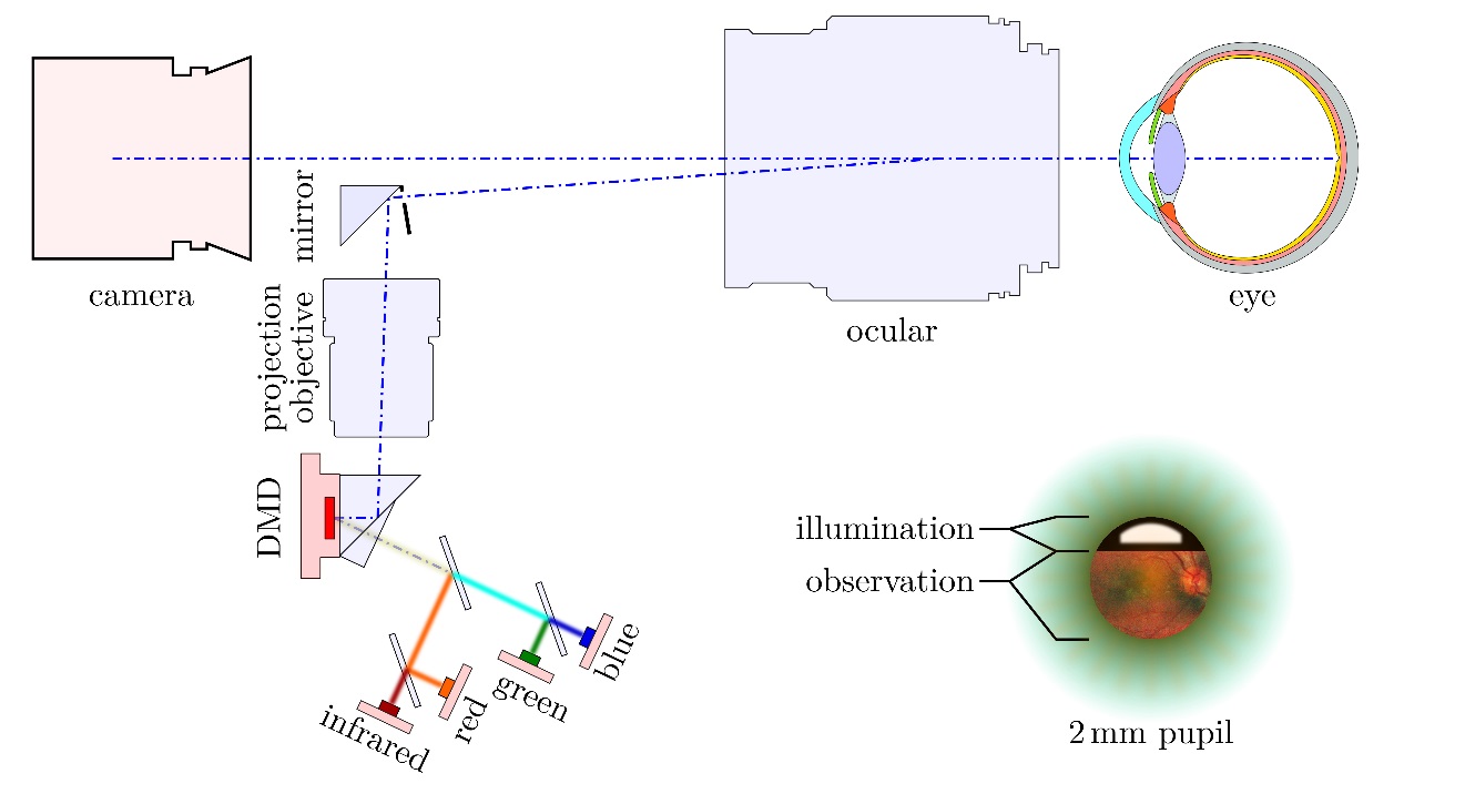
Fig 1: A DMD is integrated in the illumination path and projected onto the retina. The pupil is separatedinto a illumination area and an observation area.

Fig 2: The retina has been captured by three shots through a pupil of about 2.2 mm.
